45 fluorescent labels and light microscopy
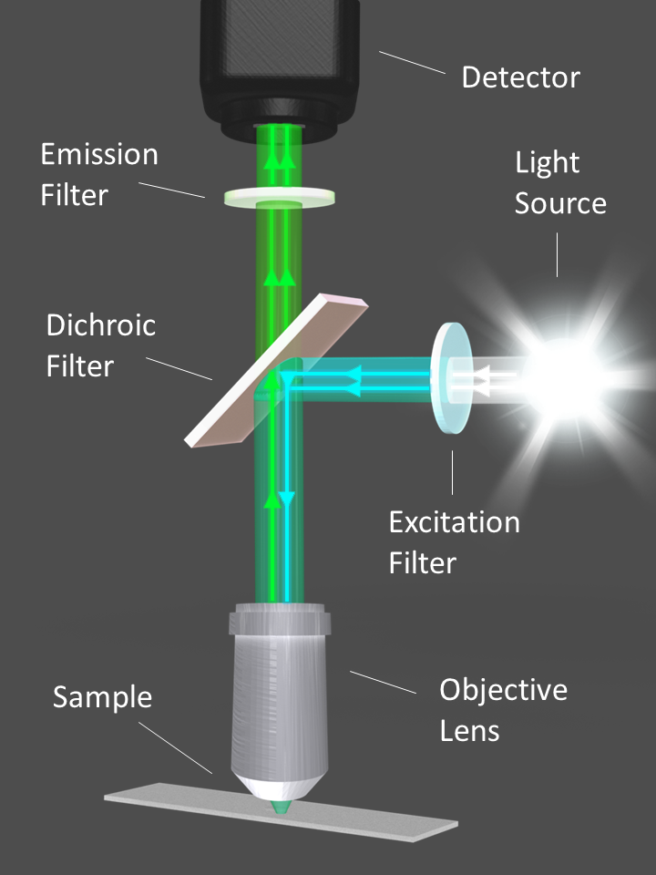
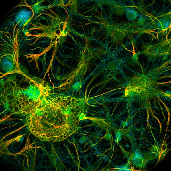
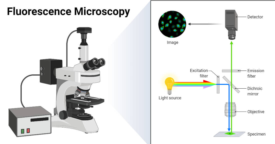
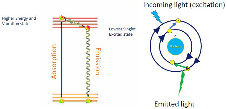
New fluorescent label provides a clearer picture of how DNA ... A molecule of interest is labelled with a special fluorescent dye that flashes on and off like a blinking star. Unlike traditional fluorescence microscopy, which uses labels that glow constantly,... Fluorescence Imaging - Teledyne Photometrics Fluorescent molecules (known as fluorophores) are used to label samples, and fluorophores are available that emit light in virtually any color. In a fluorescent microscope, a sample is labeled with a fluorophore, and then a bright light ( excitation light) is used to illuminate the sample, which gives off fluorescence ( emission light ).
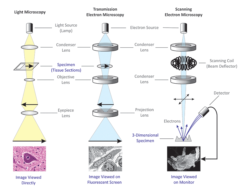
Light Microscope- Definition, Principle, Types, Parts, Labeled Diagram ... A light microscope is a biology laboratory instrument or tool, that uses visible light to detect and magnify very small objects and enlarge them. They use lenses to focus light on the specimen, magnifying it thus producing an image. The specimen is normally placed close to the microscopic lens.
Fluorescent labels and light microscopy
How do you fluorescently label mRNA for microscopy? Moffitt Cancer Center. The vendor may be able to prepare fluorescently labeled mRNA for you; fluorescently-labeled nucleotides can simply be substituted during transcription. However, you can also ... Fluorescence Microscopy - an overview | ScienceDirect Topics Fluorescence microscopy is a technique whereby fluorescent substances are examined in a microscope. It has a number of advantages over other forms of microscopy, offering high sensitivity and specificity. In fluorescence microscopy, the specimen is illuminated (excited) with light of a relatively short wavelength, usually blue or ultraviolet (UV). Using fluorescence microscopy to shed light on the mechanisms of ... Fluorescence microscopy is one of the most flexible and effective tools to characterize AMPs, particularly in its ability to measure the membrane interactions and cellular localization of peptides. Recent advances have increased the scope of research questions that can be addressed via microscopy through improving spatial and temporal resolution.
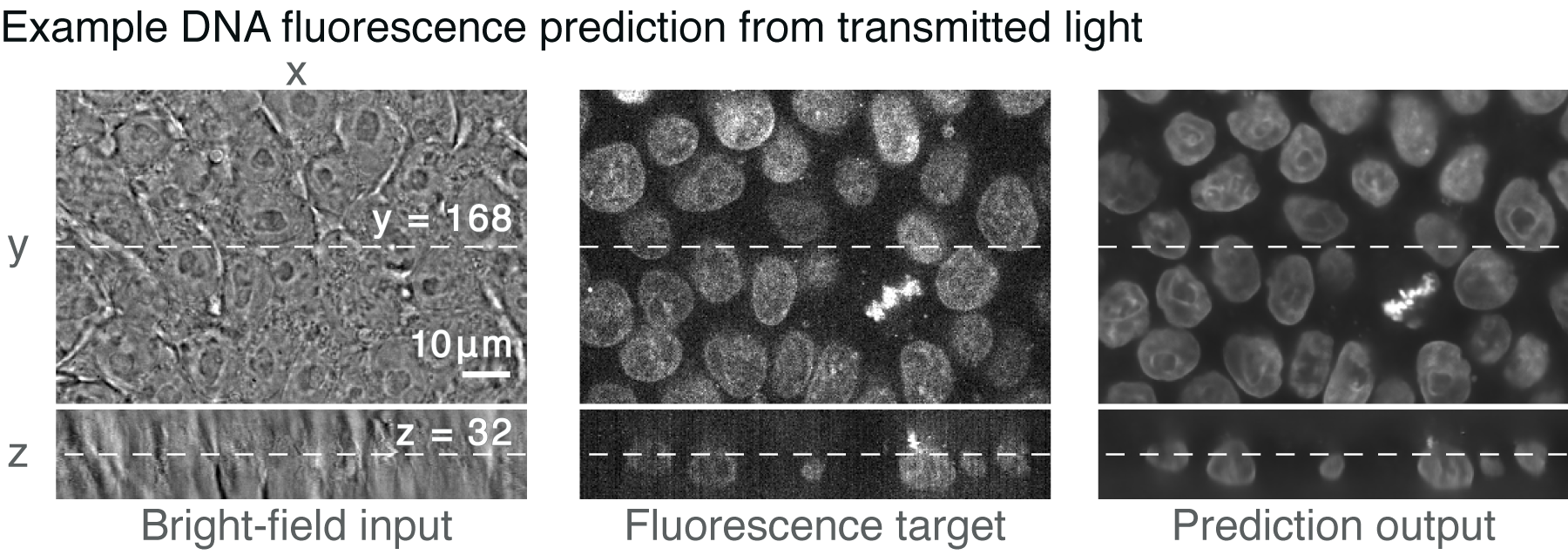
Fluorescent labels and light microscopy. Fluorescence Microscopy - Explanation and Labelled Images Fluorescence microscopy uses a high-intensity light source that excites a fluorescent molecule called a fluorophore in the sample observed. The samples are labeled with fluorophore where they absorb the high-intensity light from the source and emit a lower energy light of longer wavelength. In Silico Labeling: Predicting Fluorescent Labels in Unlabeled Images Microscopy is a central method in life sciences. Many popular methods, such as antibody labeling, are used to add physical fluorescent labels to specific cellular constituents. ... (ISL), reliably predicts some fluorescent labels from transmitted-light images of unlabeled fixed or live biological samples. ISL predicts a range of labels, such as ... Fluorescence Microscopy vs. Light Microscopy - News-Medical.net This means that fluorescent microscopy uses reflected rather than transmitted light. For example, a commonly used label is green fluorescent protein (GFP), which is excited with blue light and... Fluorescence - Wikipedia A perceptible example of fluorescence occurs when the absorbed radiation is in the ultraviolet region of the electromagnetic spectrum (invisible to the human eye), while the emitted light is in the visible region; this gives the fluorescent substance a distinct color that can only be seen when the substance has been exposed to UV light ...
Multispectral intravital microscopy for simultaneous bright-field and ... Conventional light microscopes do not allow for simultaneous bright-field and fluorescent imaging. Moreover, in conventional microscopes, only one type of fluorescent label can be observed. This study introduces multispectral intravital video microscopy, which combines bright-field and fluorescence microscopy in a standard light microscope. Different Ways to Add Fluorescent Labels - Thermo Fisher Scientific Using fluorescence provides greater contrast compared to viewing your samples with brightfield microscopy alone. Labeling various targets with separate fluorescent colors allows you to visualize different structures or proteins within a cell in the same experiment. Fluorescent Labeling - What You Should Know - PromoCell Fluorescence microscopy allows the identification of cells and cellular components and the monitoring of cell physiology with high specificity. Fluorescence microscopy separates emitted light from excitation light using optical filters. The use of two indicators also allows the simultaneous observation of different biomolecules at the same time. Light Microscopy - an overview | ScienceDirect Topics Light microscopy and electron microscopy are complementary techniques that in a correlative approach enable identification and targeting of fluorescently labeled structures in situ for three-dimensional imaging at nanometer resolution. Correlative imaging allows electron microscopic images to be positioned in a broader temporal and spatial context.
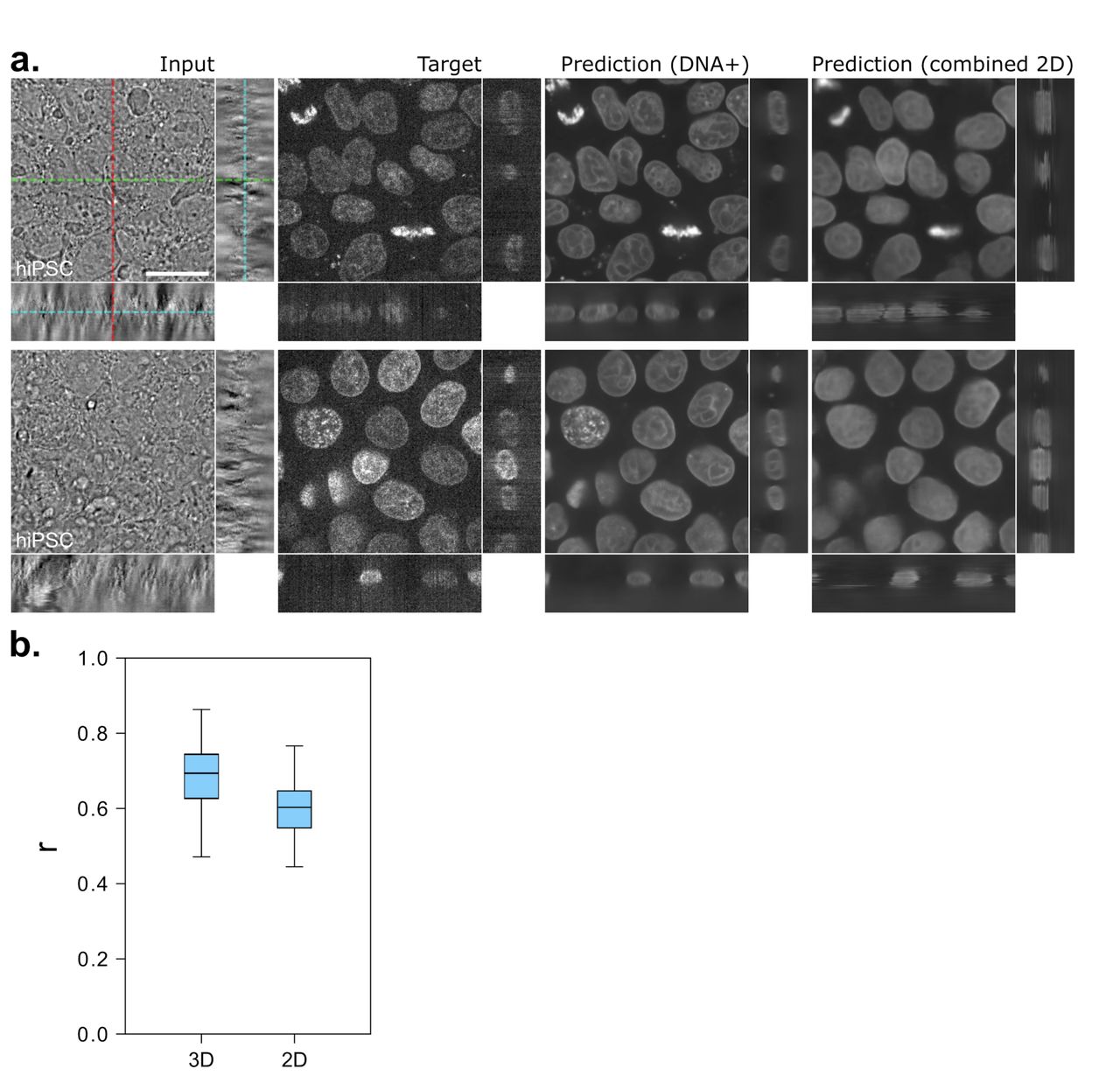
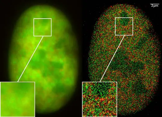
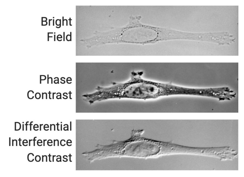
Novel Fluorescent Label Shines a Light on DNA Structure in Cancer Cells Novel Fluorescent Label Shines a Light on DNA Structure in Cancer Cells March 7, 2022 Researchers have developed a new fluorescent label that gives a clearer picture of how DNA architecture is... PDF In Silico Labeling: Predicting Fluorescent Labels in Unlabeled Images The z-stacks of transmitted-light microscopy images were acquired with different methods for enhancing contrast in unlabeled images. Several different fluorescent labels were used to generate fluorescence images and were varied between training examples; the checkerboard images indicate fluorescent labels that were not acquired for a given example. In Silico Labeling: Predicting Fluorescent Labels in Unlabeled ... - Cell The z-stacks of transmitted-light microscopy images were acquired with different methods for enhancing contrast in unlabeled images. Several different fluorescent labels were used to generate fluorescence images and were varied between training examples; the checkerboard images indicate fluorescent labels that were not acquired for a given example. Fluorescent Dyes | Science Lab | Leica Microsystems In fluorescence microscopy there are two ways to visualize your protein of interest. Either with the help of an intrinsic fluorescent signal - by genetically linking a fluorescent protein to a target protein - or with the help of fluorescently labeled antibodies that bind specifically to a protein of interest.
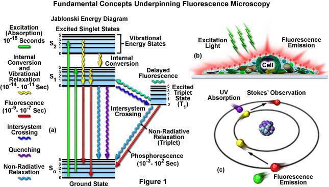
Fluorescence Resonance Energy Transfer (FRET) Microscopy Typical fluorescence microscopy techniques rely upon the absorption by a fluorophore of light at one wavelength (excitation), followed by the subsequent emission of secondary fluorescence at a longer wavelength. The excitation and emission wavelengths are often separated from each other by tens to hundreds of nanometers.
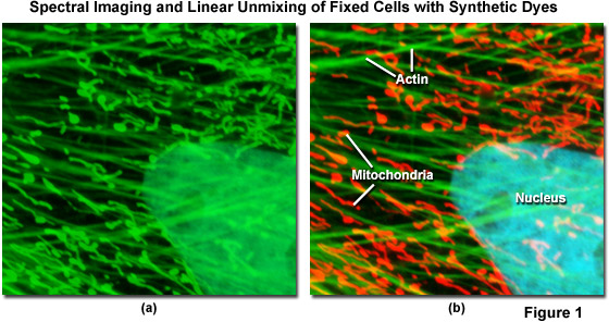
Dots, Probes and Proteins: Fluorescent Labels for Microscopy and Imaging There are currently 20 AlexaFluor dyes which span the excitation spectrum from 346 nm to 784 nm and are available as labelling kits, or conjugated to primary or secondary antibodies. Thermo Scientific produce their own probes called ' DyLight '. The ten probes which are currently available cover a spectrum from 350 nm to 800 nm.
What do I do with...? - King County - King County, Washington Sep 27, 2017 · With restrictions about COVID-19 rapidly changing, please check with individual departments to be sure a building is open before you seek in-person service.
Label-free prediction of three-dimensional fluorescence images from ... We present a label-free method for predicting three-dimensional fluorescence directly from transmitted-light images and demonstrate that it can be used to generate multi-structure, integrated...
Imaging Flies by Fluorescence Microscopy: Principles, Technologies, and ... Fluorescence microscopy in combination with specific labeling methods [ e.g., antibodies or fluorescent proteins (FPs)] enables selective visualization of the components of living matter, from molecules and organelles to cells and tissues, in both fixed and living organisms, and with high signal-to-noise ratio (SNR).
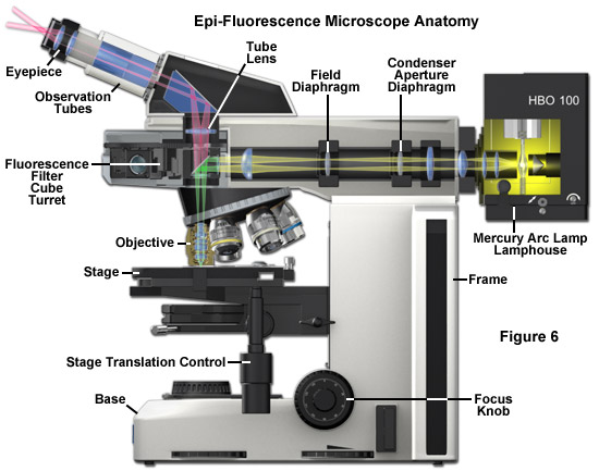
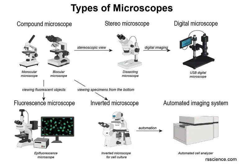
Fluorescent Microscopy A fluorescence microscope is much the same as a conventional light microscope with added features to enhance its capabilities. The conventional microscope uses visible light (400-700 nanometers) to illuminate and produce a magnified image of a sample. A fluorescence microscope, on the other hand, uses a much higher intensity light source which ...
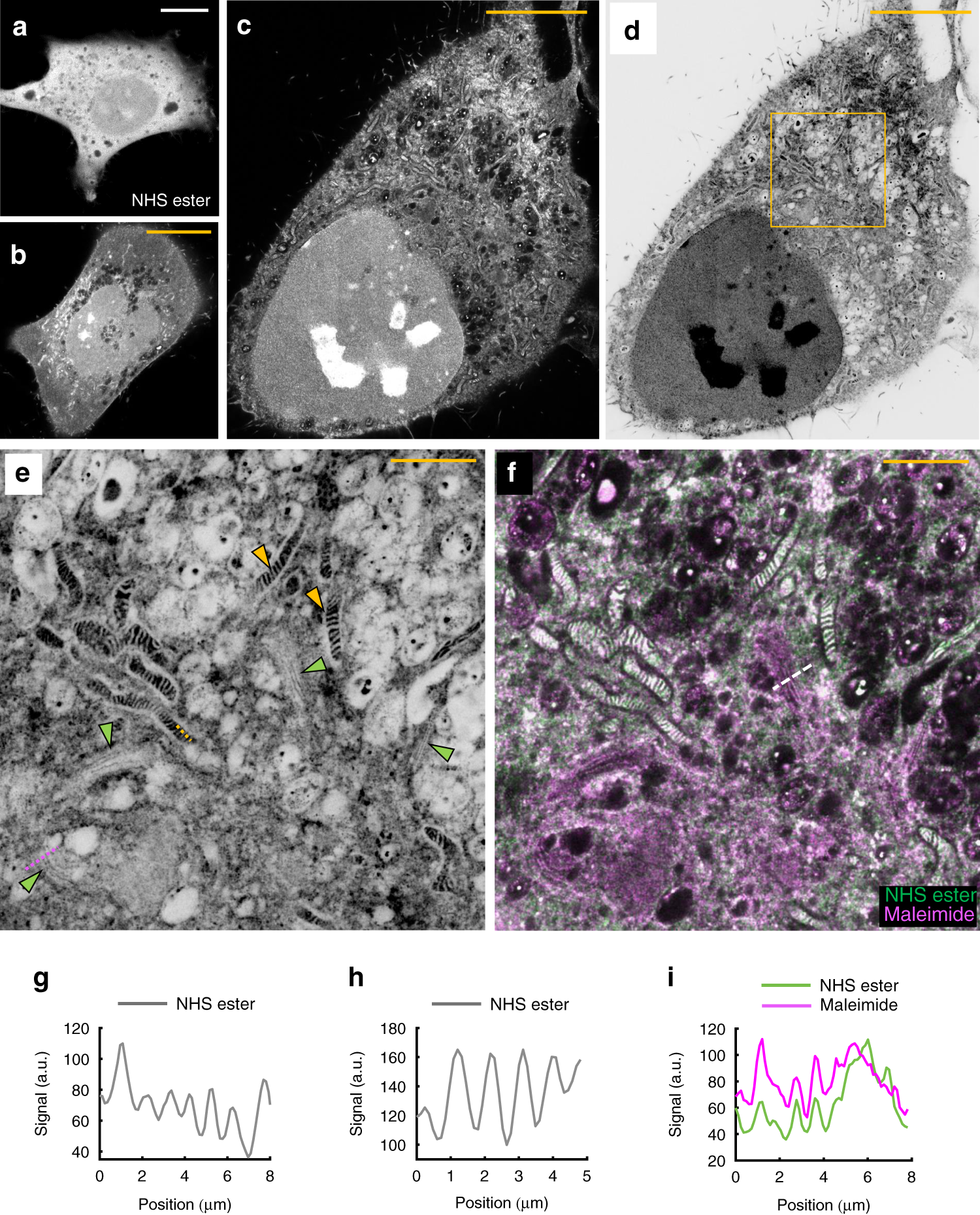
Fluorescent labeling of abundant reactive entities (FLARE) for cleared ... Fluorescence microscopy is a technique that is commonly used in the biomedical sciences. It offers the powerful ability to visualize structures or molecules in three dimensions within biological...
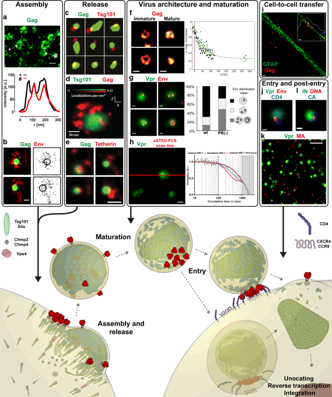
Fluorescence Microscopy & Cell Imaging | Research | UNM Cancer Center Fluorescence microscopy is routinely used to determine spatial and topological information about cells and tissues. Sophisticated laser scanning microscopic instrumentation, ultra sensitive digital cameras and specialized fluorescence probes make it possible to visualize cellular events in real time down to the molecular level.
Introduction to Fluorescent Proteins | Nikon’s MicroscopyU The brightness and fluorescence emission spectrum of enhanced yellow fluorescent protein combine to make this probe an excellent candidate for multicolor imaging experiments in fluorescence microscopy. Enhanced yellow fluorescent protein is also useful for energy transfer experiments when paired with enhanced cyan fluorescent protein (ECFP) or ...
Caveat fluorophore: an insiders’ guide to small-molecule ... Dec 23, 2021 · From its inception, fluorescence microscopy has been driven by advances in dyes. Early protein labels excited by ultraviolet (UV) light proved difficult to use for cellular imaging, and so Coons ...
Fluorescent Dyes Types, Vs Proteins, Applications Etc. Fluorescent dyes (also known as fluorophores/reactive dyes) may simply be described as molecules (non-protein in nature) that, in microscopy, achieve their function by absorbing light at a given wavelength and re-emitting it at a longer wavelength. This produces fluorescence of different colors that can be visualized and analyzed.
Labeling the ER for Light and Fluorescence Microscopy Most of them are not 100% specific for the ER membrane and may label other organelles at varying concentrations and incubation times. ... C., Wang, P., Kriechbaumer, V. (2018). Labeling the ER for Light and Fluorescence Microscopy. In: Hawes, C., Kriechbaumer, V. (eds) The Plant Endoplasmic Reticulum . Methods in Molecular Biology, vol 1691 ...
Light Sheet Fluorescence Microscopy Applications for Multicellular ... Light-sheet fluorescence microscopy has been shown to fit in this technological niche as it provides high optical resolution with 3D-imaging capabilities in large samples beyond several millimeters and up to several centimeters, while allowing the use of regular fluorescent labels or fusion proteins.
Fluorescence microscopy: established and emerging methods ... - PubMed The primary concern in all forms of microscopy is the generation of contrast; for fluorescence microscopy contrast can be thought of as the difference in intensity between the cell and background, the signal-to-noise ratio. High information-content images can be formed by enhancing the signal, suppressing the noise, or both.
Fluorescence Microscope: Principle, Types, Applications Fluorescence microscopy is a light microscope that works on the principle of fluorescence. A substance is said to be fluorescent when it absorbs the energy of invisible shorter wavelength radiation (such as UV light) and emits longer wavelength radiation of visible light (such as green or red light).
Fluorescent tag - Wikipedia S. cerevisiae septins revealed with fluorescent microscopy utilizing fluorescent labeling In molecular biology and biotechnology, a fluorescent tag, also known as a fluorescent label or fluorescent probe, is a molecule that is attached chemically to aid in the detection of a biomolecule such as a protein, antibody, or amino acid.
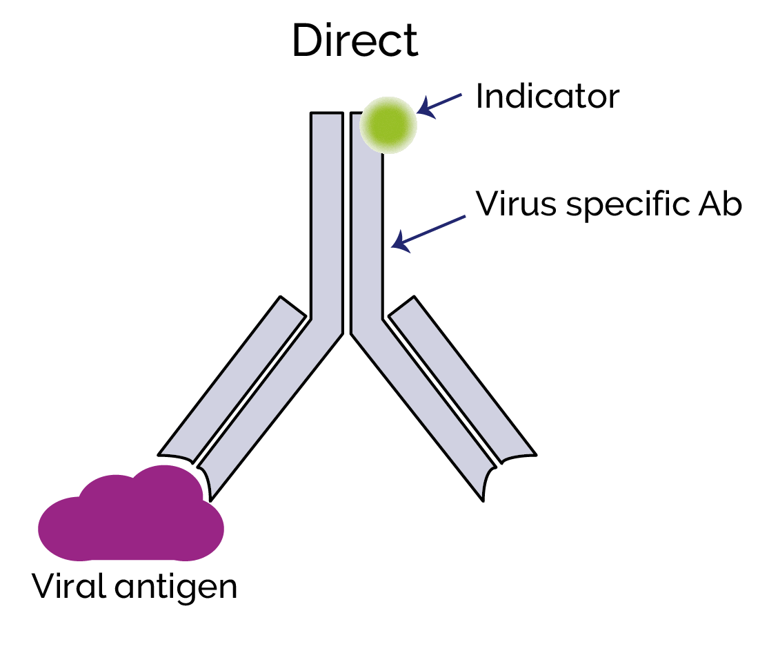
Immunofluorescence - Wikipedia Immunofluorescence is a technique used for light microscopy with a fluorescence microscope and is used primarily on microbiological samples. This technique uses the specificity of antibodies to their antigen to target fluorescent dyes to specific biomolecule targets within a cell, and therefore allows visualization of the distribution of the target molecule through the sample.
ZEISS LSM 900 with Airyscan 2 ZEN microscopy software puts a wealth of helpers at your command to achieve reproducible results in the shortest possible time. AI Sample Finder helps you quickly find regions of interest, leaving more time for experiments. Smart Setup supports you in applying best imaging settings for your fluorescent labels.
Using fluorescence microscopy to shed light on the mechanisms of ... Fluorescence microscopy is one of the most flexible and effective tools to characterize AMPs, particularly in its ability to measure the membrane interactions and cellular localization of peptides. Recent advances have increased the scope of research questions that can be addressed via microscopy through improving spatial and temporal resolution.
Fluorescence Microscopy - an overview | ScienceDirect Topics Fluorescence microscopy is a technique whereby fluorescent substances are examined in a microscope. It has a number of advantages over other forms of microscopy, offering high sensitivity and specificity. In fluorescence microscopy, the specimen is illuminated (excited) with light of a relatively short wavelength, usually blue or ultraviolet (UV).
How do you fluorescently label mRNA for microscopy? Moffitt Cancer Center. The vendor may be able to prepare fluorescently labeled mRNA for you; fluorescently-labeled nucleotides can simply be substituted during transcription. However, you can also ...














Post a Comment for "45 fluorescent labels and light microscopy"